Image Core News
Image Core News
* December 14, 2020: Current JHU space occupancy limit is 1 person per 200 square feet (one person per small lab or office - limit for each room is posted at each door). Please wear your face mask, wash hands frequently, bring your own lab gloves for touching microscope, tables, keyboards, computer mice. First covid-19 vaccine arriving this week for clinical staff, likely shortage for several months, so plan for covid=19 restrictions to continue in our image core space through at least April 2021 and possibly July 2021 (and potentially beyond if vaccine protection wanes in some people).
* September 2020: We won! Lauren Blake (PhD student, Bin Wu's lab) and GM won 1st prize of Lumencor's 2020 Earthday competition. $10,000 list price Lumencor SOLA SE U-nIR lamp.
* June 15, 2020 - image core covid-19 re-opening phase 1. All users must read and abide by our re-opening policies http://confocal.jhu.edu/covid-19-policies
* March 27, 2020 - Leica is offering 90 day "Leica LAS X at Home" - free (just need to register with your work email address - all JHU employees can access "web Outlook" through MyJHU), link is
https://www.leica-microsystems.com/las-x-home-office-license/
Note: ~1.2 Gigabyte download.
You can upload up to 5 Terabytes on your JHU Microsoft OneDrive (myJHU --> Cloud --> OneDrive).
I emailed our Olympus reps encouraging them to top Leica byh providing fully loaded cellSens AND FluoView free for 90 days ... and the mysterious NoviSight software too.
***
The Ross Fluorescence Imaging Core site is under new management (May 2017). John Gibas is now with Olympus. George McNamara, PhD, is now the image core manager. Prof. Olga Kovbasnjuk has moved to University of New Mexico. Prof. Bin Wu is now the image core Director.
There will be more content and changes coming to these pages in the near future. All users should provide JHU IO Numbers (account numbers) for billing (11/2019: now all reservations and billing is done through iLab ... sessions are rounded up in 30 min interval based on the required sign-in sheet times).
George McNamara office: Ross Bldg S913 (S = Service corridor).
Instruments: Ross S913, S910, S972.
Data: usually save to acquisition PC (D: or fastest drive as designated by GM), transfer to user's Microsoft OneDrive account (MyJHU --> Cloud --> Microsoft OneDrive). Every JHU user gets 5 TB OneDrive space, and can share their OneDrive data with their PI.
//
Our major equipment - web links - descriptions and iLab schedulers
==>iLab links (should) work if you have signed in to iLab and have access to the instrument in iLab:
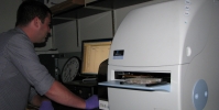
Olympus FV3000RS Confocal Microscope
FV3000RS iLab https://johnshopkins.corefacilities.org/schedules/447115#/schedule
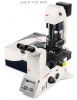
Leica SP8 Confocal Microscope (owned by ACCM, managed by us)
SP8 iLab https://johnshopkins.corefacilities.org/schedules/345585#/schedule
FISHscope (new - limited access, here because very cool)
FISHscope iLab https://johnshopkins.corefacilities.org/schedules/447181#/schedule
All Current Equipment (note: may break onto 2 pages)
iLab for All Ross FIC Current Equipment (all but ACCM Leica SP8)
https://johnshopkins.corefacilities.org/sc/5148/ross-imaging-center/?tab=equipment
iLab reservations require JHU IO# be assigned by the PI to the user. See
Agilent iLab support page for membership requests and fund numbers
for how to do this (most departments or Dept of Medicine divisions financial offices can help - if need more help, email Shawn Franckowiak (JHU SOM Research Dean office) and Jeffrey Smith (Cancer Center office).
//
Monday, November 4, 2019
1. Ross Imaging Center went live on iLab today, URL is
https://johnshopkins.corefacilities.org/sc/5148/ross-imaging-center/?tab=equipment
Trained users will need a valid JHU account (aka IO#) to make reservations. New users should contact Dr. George McNamara, core manager, about training. For the Fv3000RS and SP8 confocal microscope you can expect to be charged $127/hr for four hours (usually two 2 hour sessions -- assumes you have research level light microscopy experience and need training with respect to our confocal microscopes). This is $127 = $100 trainer's time + $27/hr instrument time. We do not do classroom training - you can get that through Prof. Scot Kuo, MicFac, https://microscopy.jhmi.edu/index.htm ... if you have little or no knowledge of light microscopy, you should tell us in advance, and be prepared t pay for additional time to get training (10 hours at $127/hr may or not be enough). You can also consider starting on one (or more) of our basic microscopes and use that.
Please note that the Leica SP8 confocal microscope is in the ACCM Confocal Microscope Core (we manage the SP8 for ACCM).
Our Conte Center web site has been updated
Center http://jhugicc.org
Image Core http://jhugicc.org/cores/core-b-imaging
//
September 18, 2019 news
Schedulers are moving to Agilent iLab Organizer ("iLab") in October 2019 ("go live" expected Mon Nov 4, 2019). We have been using Google Calendars for a couple of months since this web site's scheduler hosting had issues.
Data transfer:
1. Please "save locally" (on core PC's that have data drives -- do not save any data on any of our C: drives and NEVER use a USB stick or portable drive on any image core PC ... these are subject to confiscation and being tossed in the biosafety trash) and , and upload to your MyJHU -> Cloud -> Microsoft OneDrive (once upload is done, lgout of OneDrive and then myJHU). many of our PC's now have local 10Gbe Ethernet and ASUS Hyper M.2 PCIe fast data drives. We are working on connectign to the JHU I.T. network at 10 Gbe to improve upload speed to user's OneDrive.
Acces to our file server:
==> Note: please do not publicize the name of our file server.
If you have a compelling reason to use use our file server, and lose access because your computer is now on a JHU SOM domain, you may be able to connect using Windows Map Network Drive, by selecting "Connect using different credentials" and appropriate server name and path ... you may need an account on our server (hint: use JHU's Microsoft OneDrive instead, please).
//
August 6, 2019 news:
FISHscope now operational (ok, not quite: we had a loaner Lumencor SPECTRA X from Olympus, that demo period ended, we are waiting for our Lumencor SPECTRA III-360 to arrive) ... currently restricted use to the single molecule RNA FISH the microscope was purchased for.
Andor Revolution X1 spinning disk confocal microscope - now Legacy instrument - operational for existing fully trained users, if laser(s) etc fail, not planning on replacing components. Has MetaMorph Premier, so can be used as an additional offline workstation (few users use Pemier features).
Image Core usage: FV3000RS, ACCM Leica SP8, Keyence BZ-X710 "all-in-one" microoscopes very heavy use.
//
June 1, 2018 the ACCM Leica SP8 confocal microscope we manage is on iLab
https://johnshopkins.corefacilities.org/service_center/3804/
//
July 20, 2018: Zeiss LSM510META confocal microscope has been retired.
//
August 3, 2018 update:
Olympus FV3000RS confocal microscope has been funded by NIH S10 grant proposal. we expect to have 'up and using' by end of August 2018.
FISHscope expected to be funded (NIH P30 G.I. Center supplement plus matching funds from us) (really: smFISHscope). Microscope delivery asnticipated "autumn 2018".
G.I. single molecule fluorescence in situ hybridization (smFISH) first 30 G.I. centric probe sets development and implementation funded.
Instructions to (hopefully) see our file server is now on our McTips page
http://confocal.jhu.edu/mctips
(GM can also send as a Word doc).
//
August, 2018: Olympus FV3000RS confocal microscope is here!
November 2018: The NIR unit arrived, now have:
7 laser lines: 405, 445, 488, 514, 561,640, 730 nm.
6 detectors:
* 4 GaAsP (internal, spectral bandpasses, 400-800nm)
* 2 GaAs (external, NIR1: 690-750nm, NIR2: 780LP; conventional IX83 filter cube, so could buy additional cubes for specialty needs). We would love a contribution to get additional external detectors!
--> corssover point of sensitivity of GaAsP (better vis) and GaAs (better NIR) is ~712 nm.
March 2019: FV3000RS ZDC 830 nm installed (previously was shorter wavelength). ZDC = Zero Drift Compensation.
April 22, 2019: FISHscope has been ordered. Main use will be single molecule RNA fluorescence in situ hybridization (smFISH).
* Olympus IX83 inverted microscope ('gliding' stage, motorized Z, 2nd deck is Sutter 10-3 emission filter wheel).
* Lumencor SPECTRA 3-360 lamp (8 LEDs, 360 nm to 750 nm).
* Semrock Penta filter cube (penta exciter, penta dichroic,penta emission in cube) and emission filters for Sutter wheel.
* Hamamatus ORCA-FLASH4.0LT.
* LCI optogenetic unit for screening.
* Generously funded by our NIH P30 G.I. Conte Center grant supplement and internal funds.
//
gmcnama2@jhmi.edu
geomcnamara@earthlink.net