ACCM_IncuCyte_S3

Live cell imaging system inside 37 C incubator - short to long term (many days possible) live cell imaging flasks and SBS plates (can hold up to 6)
Location: Ross3
ACCM Incucyte S3 Live-Cell Imaging and Analysis System
Live cell imaging system inside 37 C incubator - short to long term (many days possible) live cell imaging SBS plates (can hold up to 6 plates - has inserts for imaging dishes) ... our unit does not work with flasks.
iLab web site for ACCM core: https://johnshopkins.ilab.agilent.com/service_center/3804/?tab=equipment
iLab weekly calendar page for IncuCyte S3: https://johnshopkins.ilab.agilent.com/schedules/519999#/schedule
* Rate is $2/hr (this may be revised to be per well). Available for fully trained users of this facility (if you were trained on an Incucyte, you still need Biosafety etc training for this unit, along with access information for the data).
* 6 position imaging system inside 37 C, 5% CO2 incubator. Each position can hold an SBS plate (ex. 6, 12, 24, 48, 96-well plate). Lid on or cap loose. ... again: our unit does not have a holder for flasks.
* Biosafety: Do NOT contaminate the Incucyte, incubator or room with bacteria, fungi, viruses, HeLa or other cell lines. Expect to be charged for clean-up time. If your experiment involves deliberate infection of cells get clearance from the instrument host: Prof. Nicola Heller <nheller@jhmi.edu>
* Set up/set off would be during typical work hours 9am-5pm weekdays. Set by staff.
* iLab requires the user to have a valid Cost Object (aka I/O aka account number).
Enables single timepoint, few hours, many hours, optionally many DAYS timelapse imaging. You can search Google Scholar, ScienceDirect, Highwire Press, bioRxiv, etc, for publications / preprints that use Incucyte with your favorite cells etc.
One of the original (maybe the original) use was to "keep an eye" (camera) on cells growing in SBS plates to (i) check that initial cell density is good, (ii) cell growth rate is good, (iii) which flasks have reach the correct point for optimal harvesting for high throughput screening (HTS) or high content screening (HCS) - which could be conducted on Incucyte and/or other instruments.
Description from https://www.sartorius.com/en/products/live-cell-imaging-analysis/live-cell-analysis-instruments/s3-live-cell-analysis-instrument
Incucyte S3 Live-Cell Imaging and Analysis System
With the Incucyte® S3 Live-Cell Analysis System, take your research further with automatic acquisition and analysis of cells in a physiologically relevant environment. See more information in every sample and explore more applications - from cell health to complex functional assays - through utilization of up to two total fluorescence and HD phase imaging channels simultaneously.
Key Features
Robust application suite
Multi-user, multi-application support through remote network capability and unlimited user licenses
Two-color Green|Red plus HD phase contrast imaging
4X, 10X, 20X objectives on an automated turret
Support for three interchangeable vessel trays and over 700 vessels, compatible with dishes, slides and SBS plates - up to six SBS plates in parallel.
Analyze even the most sensitive living cells around the clock for days, weeks or months. Never miss powerful insights again with the Incucyte® S3 Live-Cell Analysis System and associated reagents, consumables and software!
****
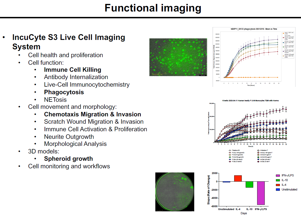
Examples od Incucyte assays (a few highlighted)
IncuCyte S3 Live Cell Imaging
System
• Cell health and proliferation
• Cell function:
• Immune Cell Killing
• Antibody Internalization
• Live-Cell Immunocytochemistry
• Phagocytosis
• NETosis
• Cell movement and morphology:
• Chemotaxis Migration & Invasion
• Scratch Wound Migration & Invasion
• Immune Cell Activation & Proliferation
• Neurite Outgrowth
• Morphological Analysis
• 3D models:
• Spheroid growth
• Cell monitoring and workflows
Visual:

Reserve Equipment

